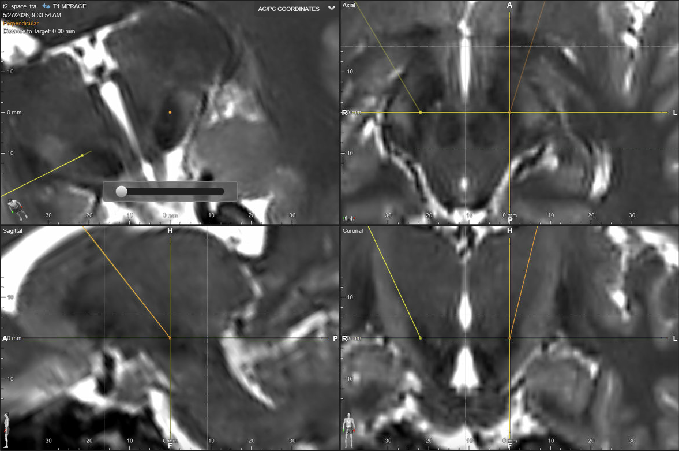
Four-panel DBS planning screenshot for STN targeting showing probe-eye, axial, sagittal, and coronal views of the planned trajectory through the subthalamic target region.
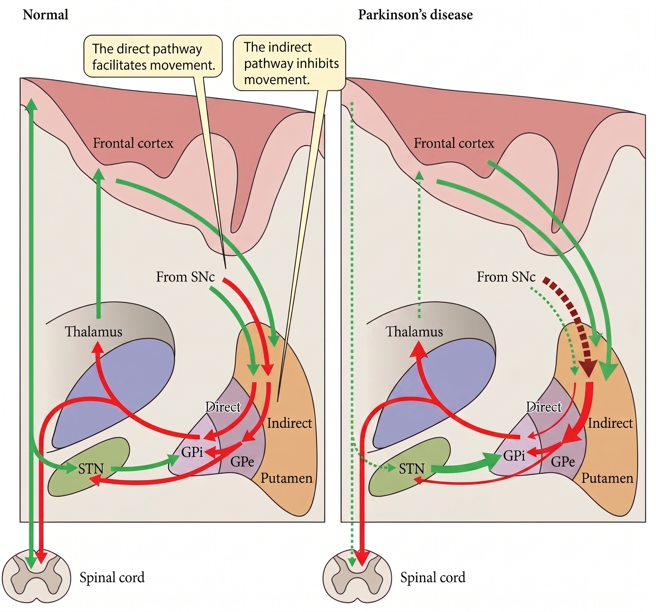
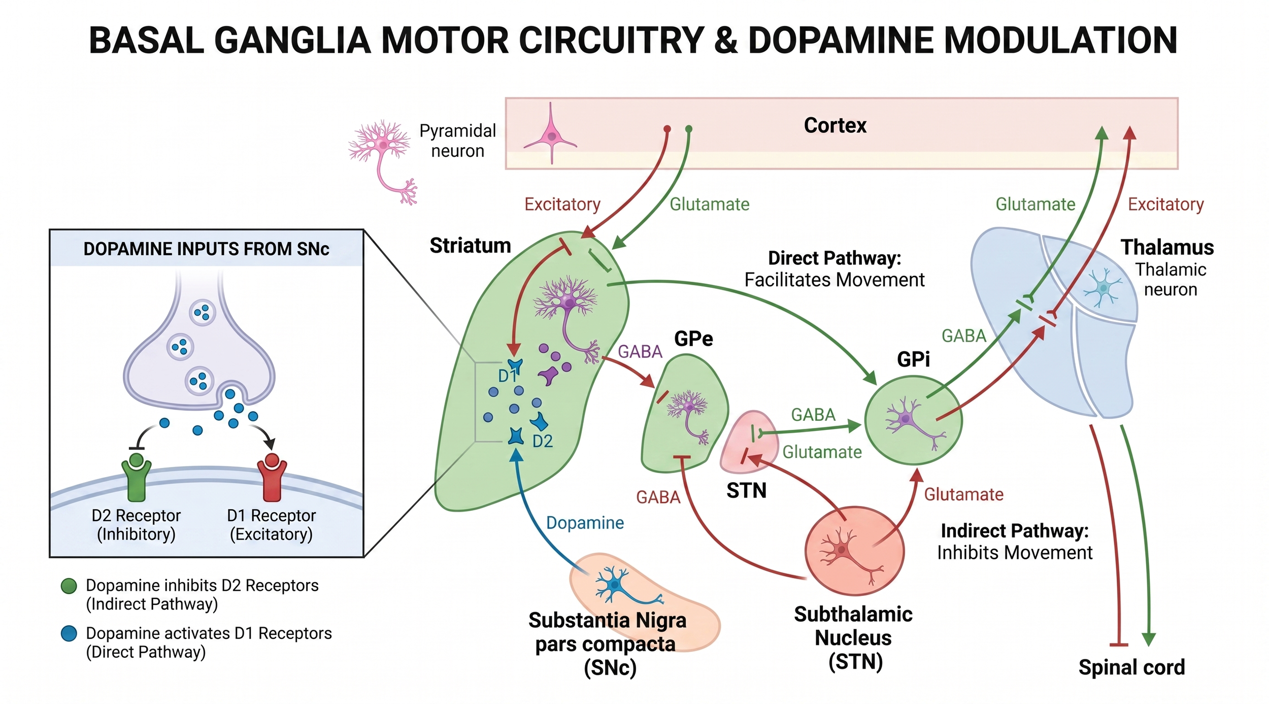
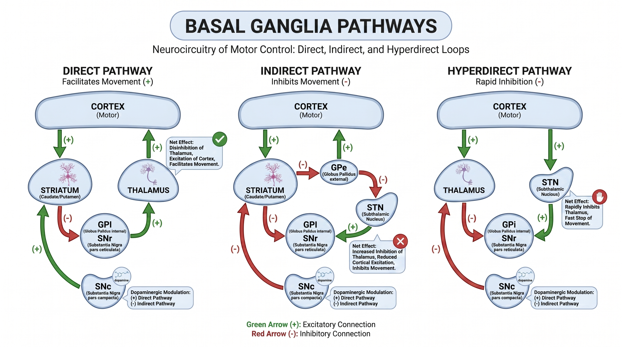
Diagram of the Albin-DeLong model of the basal ganglia direct and indirect pathways
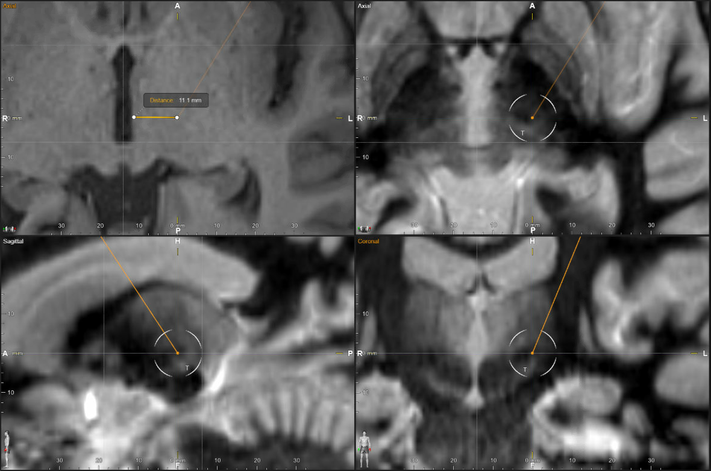
Four-panel DBS planning screenshot for Vim targeting showing axial T1 lateral-wall measurement, axial FGATIR target review, and sagittal and coronal FGATIR trajectory confirmation.
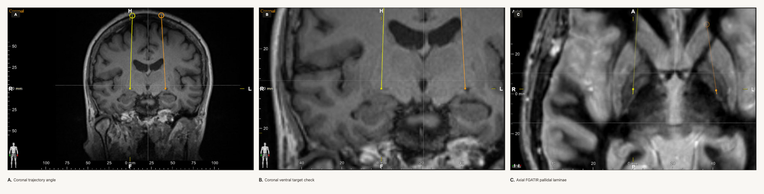
Three-panel DBS planning screenshot for GPi targeting showing coronal lead angle, close coronal ventral target position near the optic tract region, and axial FGATIR pallidal laminae with lead position.
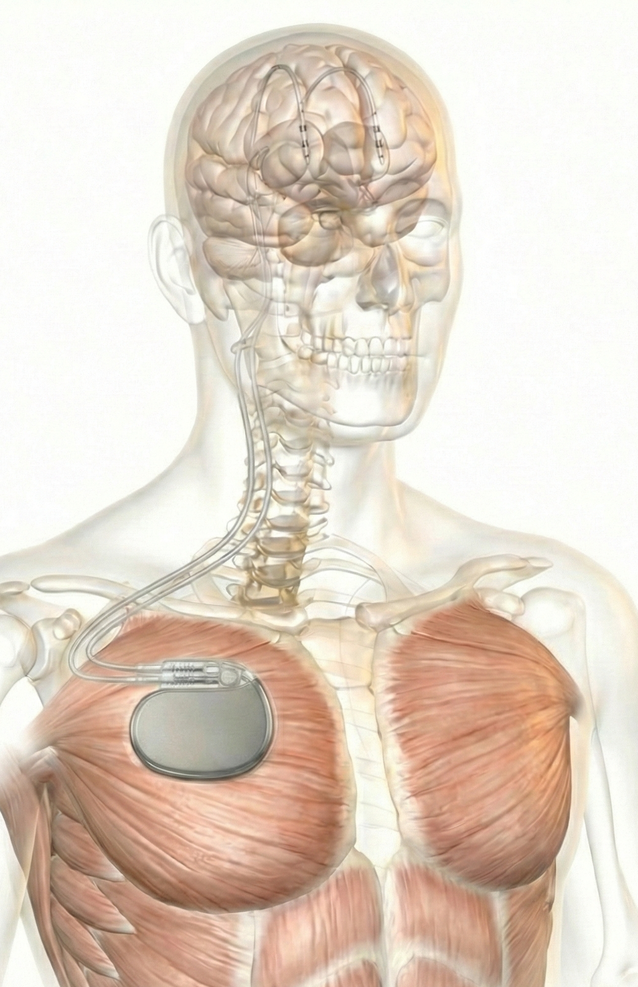
Transparent anatomical illustration of bilateral deep brain stimulation leads implanted in the brain and connected to an implanted pulse generator in the chest.
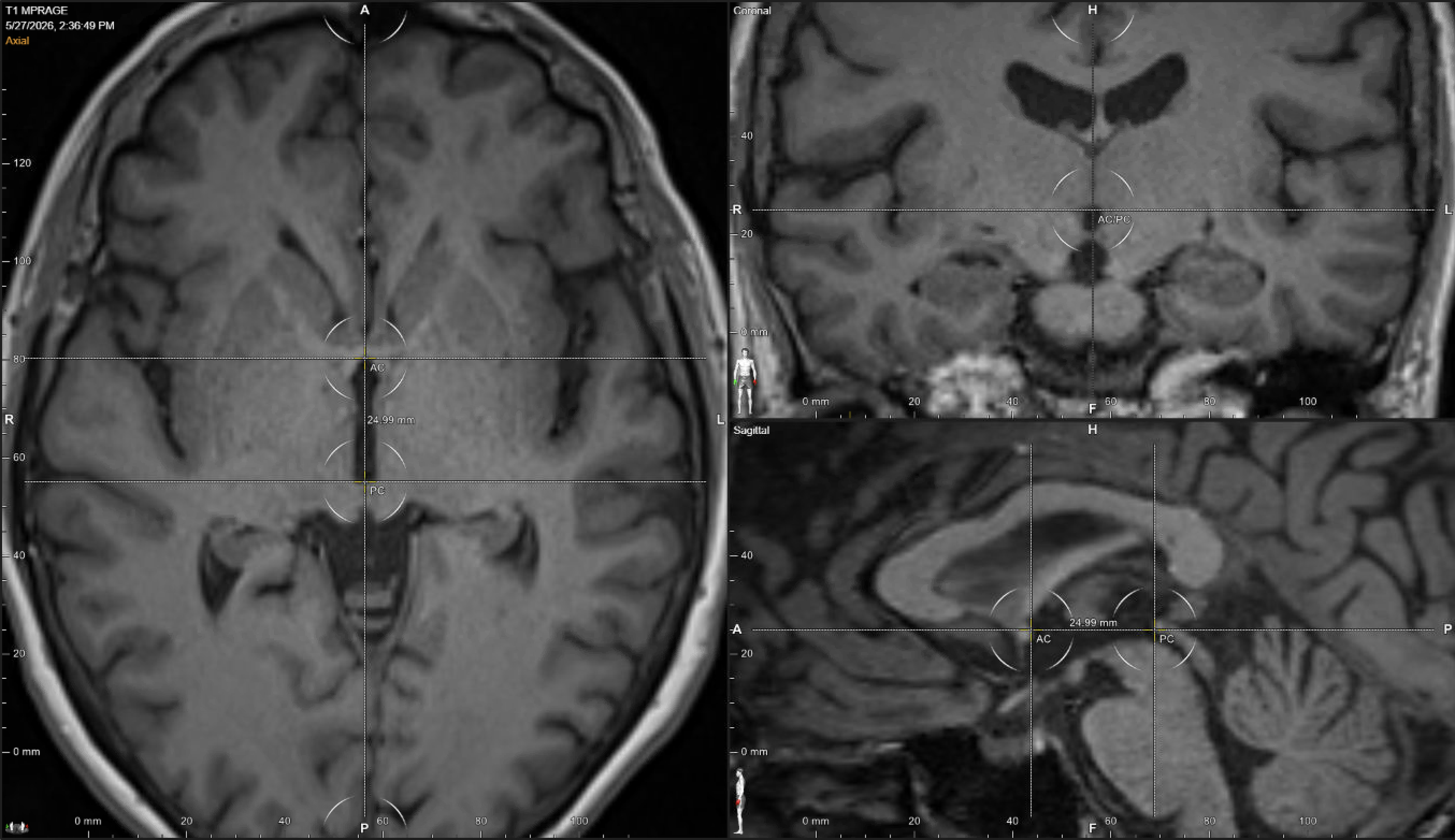
Three-panel DBS planning screenshot showing sagittal AC-PC landmark selection, axial intercommissural plane confirmation, and coronal midline alignment for stereotactic coordinate setup.
Diagram of basal ganglia motor circuitry
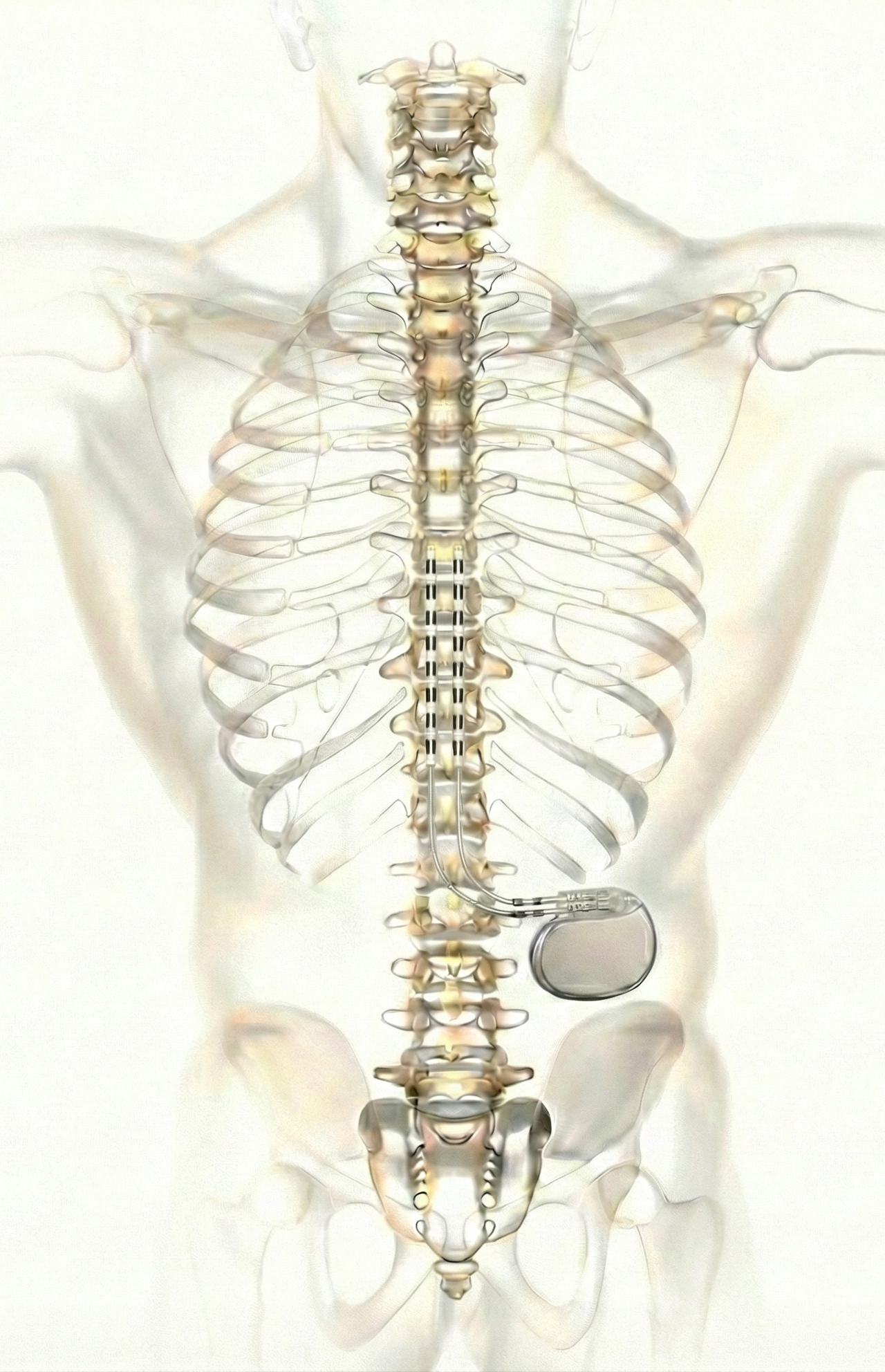
Posterior anatomical illustration of spinal cord stimulation leads positioned along the thoracic spine and connected to an implanted pulse generator in the flank.
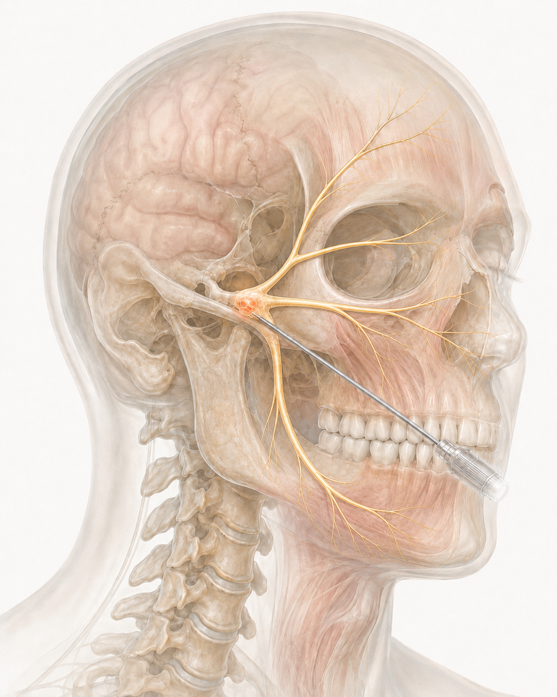
Illustration of a needle passed through the cheek toward the trigeminal ganglion for percutaneous treatment of trigeminal neuralgia
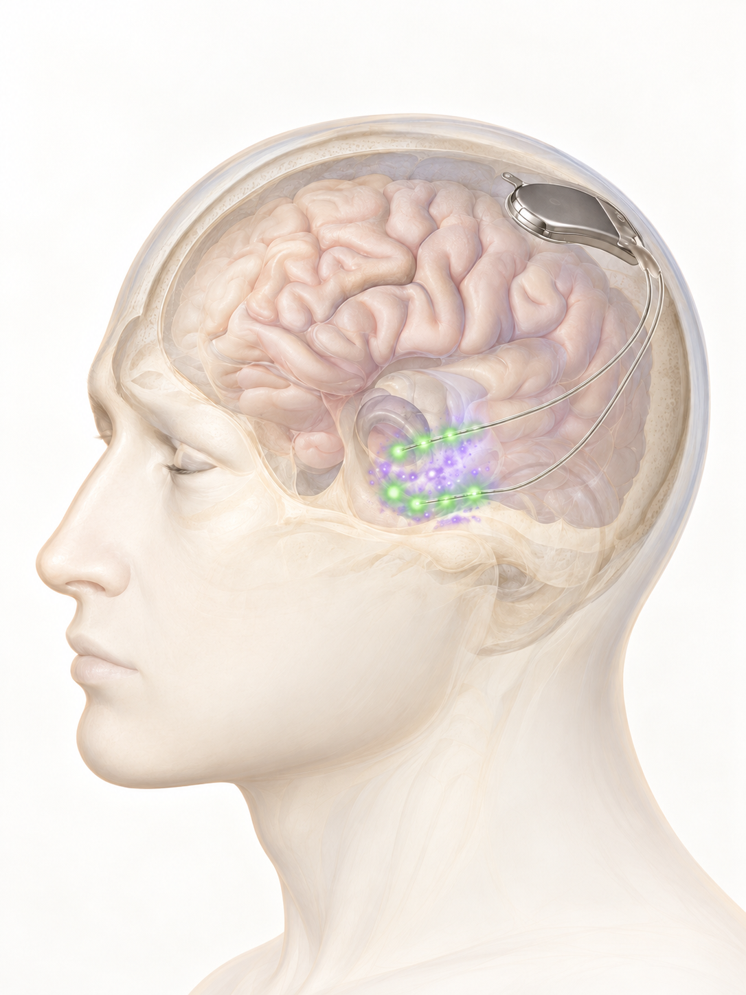
Illustration of a responsive neurostimulation device in the skull with leads placed in the brain
Instagram logo
Diagram of basal ganglia pathways
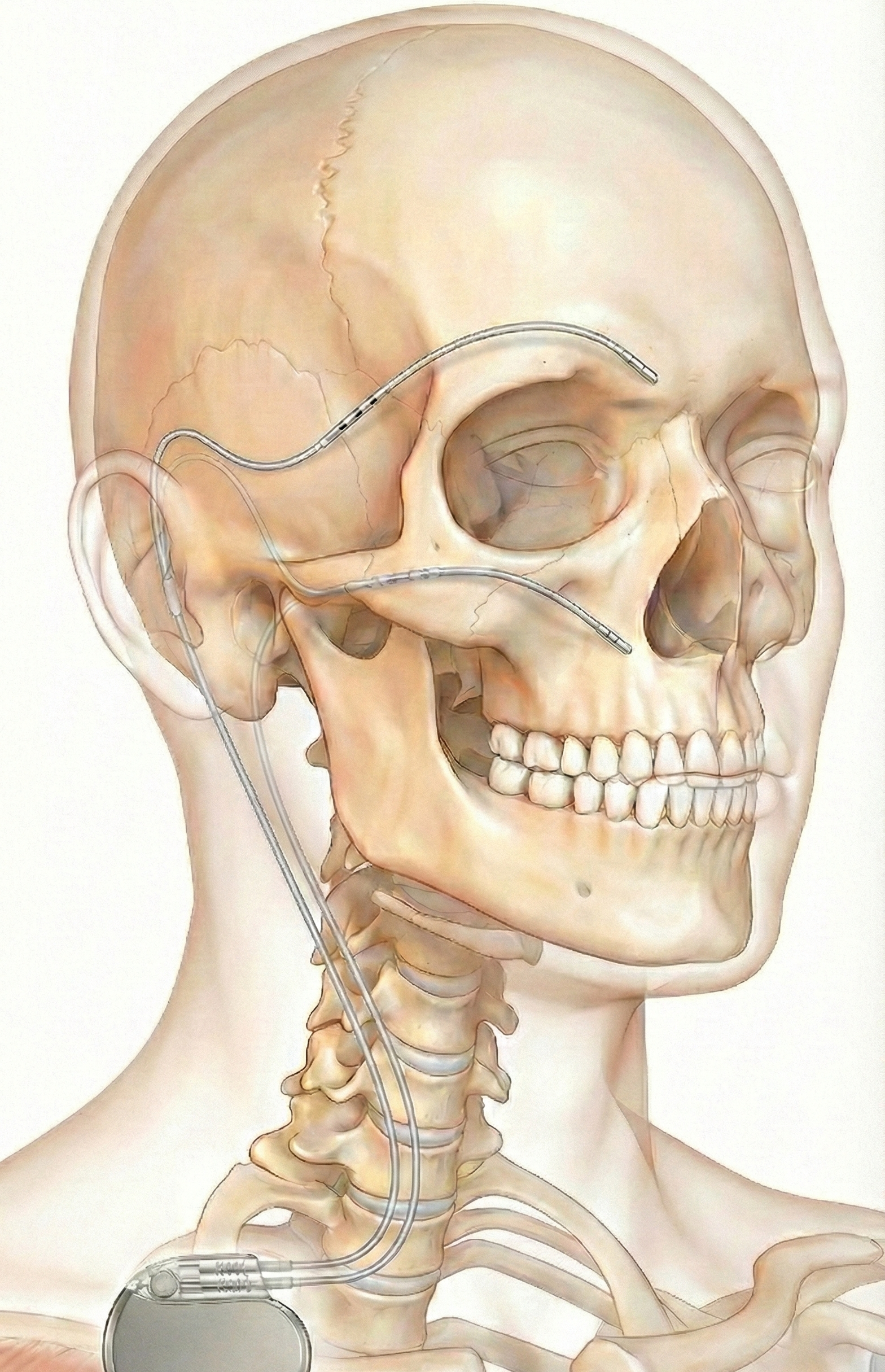
Lateral anatomical illustration showing craniofacial neuromodulation leads tunneled over the face and connected to an implanted pulse generator.
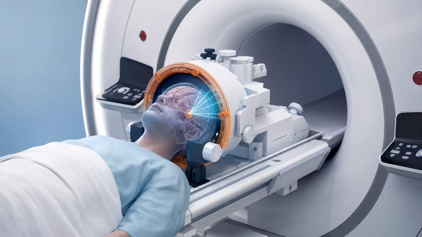
Patient in an MRI scanner wearing a focused ultrasound helmet for incisionless treatment
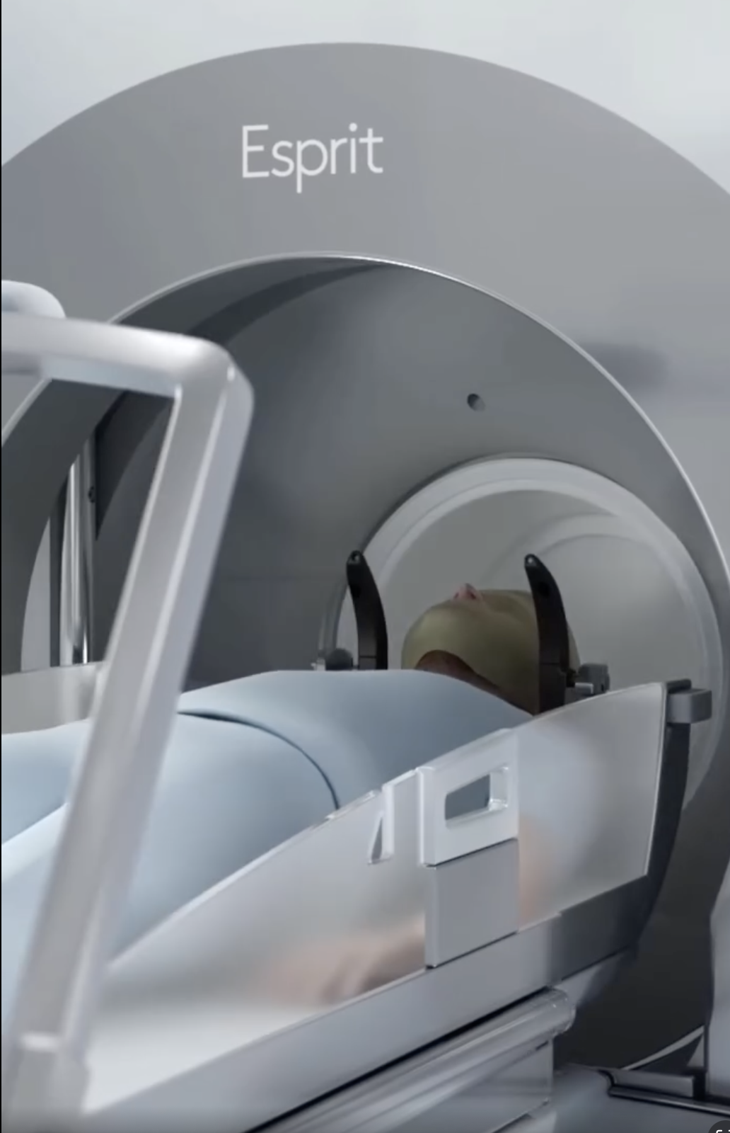
Patient positioned in an Elekta Esprit Gamma Knife radiosurgery unit with head immobilization for stereotactic treatment.
Timoteo Almeida, functional neurosurgeon, Pittsburgh.
Illustration of radiation beams converging on a target in the brain for stereotactic radiosurgery
Portrait of Dr. Timoteo Almeida